The human brain is constructed of about 100 billion individual neurons (or nerve cells). Each neuron is plays a part in making someone who they are: memories, behavior, quirks, and preferences. When a patient suffers a seizure it’s due to rapid neuronal firing. When a patient becomes unresponsive due to hypoglycemia it’s caused by lack of glucose to the neurons which make up the area of the brain keeping them conscious. Neurons are also found all throughout the body. In fact, the second largest concentration of neurons is in the gut (called the enteric nervous system), and coordinates the digestion of food. [As an aside, patients who take selective-serotonin re-uptake inhibitors, SSRIs, commonly suffer stomachache and indigestion due to the blocking of the neurotransmitter serotonin in the enteric nervous system. This is a side effect of blocking the re-uptake serotonin in the brain, thus increasing available serotonin levels, to treat psychiatric disorders]
Let’s take a closer look at a motor neuron (specialized for muscle movements):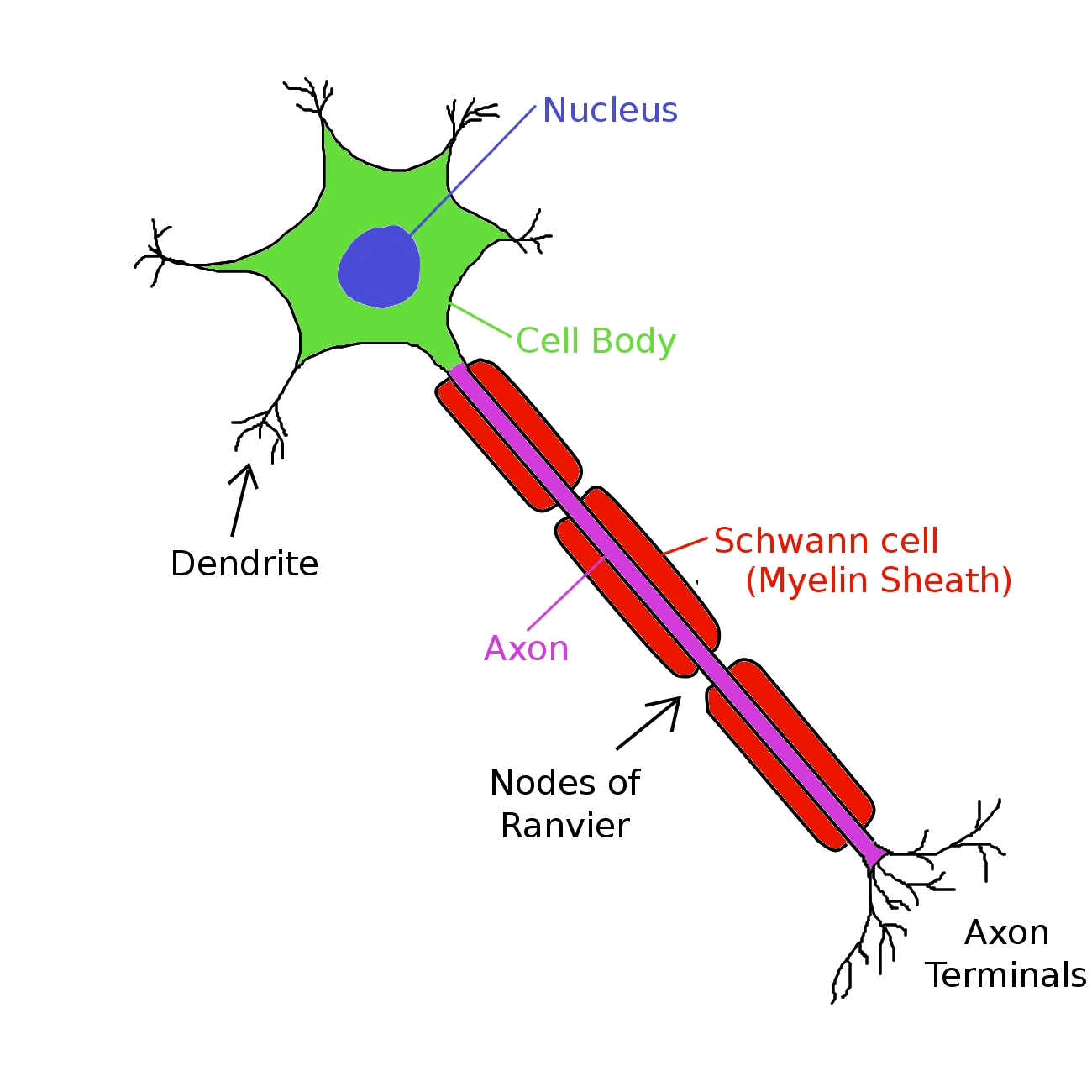
The large green area above marked as the cell body, also known as the soma, houses the nucleus. Projecting out from the cell body are dendrites which function to receive impulses, similar to antennae. The long, thin projection in purple is called an axon. Axons are specialized tosend impulses.
In red are Schwann cells, a type of glial cell. Glial cells function to support the neuron and the Schwann cells wrap around the axon and supply a layer of a lipid substance called myelin to insulate and increase the conduction speed of impulses. A number of diseases, such as Guillain-Barre and Charcot-Marie-Tooth, are related to demyelination. It’s important to note that Schwann cells are only found in the peripheral nervous system. Central nervous system cells are also myelinated, however a different type of glial cell (an oligodendrocyte) performs the myelination.
Interrupting the myelination are nodes of Ranvier which function to boost the signal strength. At the end of the axon are terminals. In the terminals are tiny sacs, called vesicles, which contain neurotransmitters.
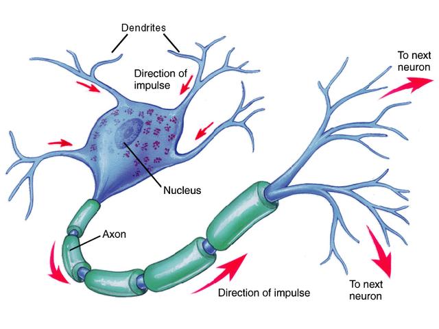
The image below traces the flow of a signal through a neuron. Remember, axons carry signals away from the nucleus and dendrites carry signals towards the nucleus. This is the general rule however in medicine there are always exceptions. Some neurons send signals via dendrites.
Each axon connects to (or innervates) another neuron or in some cases directly innervates a muscle cell or gland. The junction at which an axon connects to another cell is a synapse.
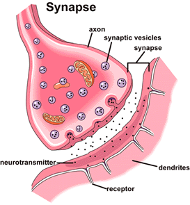
Once the neurotransmitters are released from the vesicles they cross the synapse and attach to a receptor on the other side. This allows the signal to continue (called propagation) until it reaches its’ destination. For additional, check out the link below:
Khan Academy – “Anatomy of a Neuronâ€
![]()